
More project photos (3)

The problem
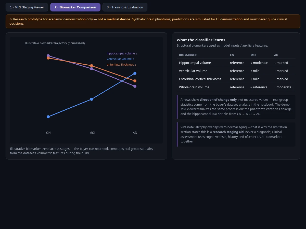
Alzheimer's disease begins damaging the brain years before memory symptoms appear, and structural MRI can reveal its fingerprints — hippocampal shrinkage, ventricular enlargement, cortical thinning — long before a clinical diagnosis. But measuring those changes by eye is subjective, and early stages like mild cognitive impairment (MCI) overlap heavily with normal aging, which is why automated staging is an active research area. The Alzheimer's Disease Neuroimaging Initiative (ADNI) has collected standardized longitudinal MRI for this exact purpose, making it the canonical academic dataset for the task. This project builds the standard student approach: a 3D CNN (with a slice-based 2D alternative) that stages scans into AD, MCI or CN, trained with strict subject-wise splitting — the methodological point examiners probe first, since random slice splits silently leak and inflate accuracy. It demonstrates neuroimaging ML end to end while being explicit that it is a research staging aid, never a diagnosis.
T1-weighted MRI scans (ADNI via approved access; OASIS 2D slices for quick-start), labeled AD, MCI or CN; splits are subject-wise.
Follow the working sequence
How it works
- T1-weighted brain MRI scans are sourced from ADNI (via approved LONI IDA access) or from the openly downloadable OASIS 2D slice set for the quick-start path, with labels AD, MCI or CN.
- Volumes are skull-stripped, registered to a common template, intensity-normalized and resampled; the 2D path extracts mid-temporal axial slices.
- Subjects — never individual slices — are split into train/validation/test sets, so no brain appears in more than one split.
- A 3D CNN (or 2D EfficientNet on slices with majority voting per subject) trains with augmentation — flips, rotations, elastic deformations — logging loss and per-class metrics per epoch.
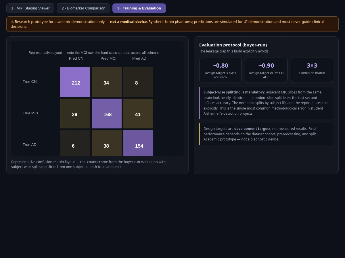
- The evaluation notebook computes final metrics on the held-out subjects: 3-class accuracy, per-class precision/recall/F1, 3x3 confusion matrix and AD-vs-CN ROC-AUC, plus Grad-CAM volumes.
- The Flask demo stages an uploaded scan, displays per-class probabilities with the hippocampal ROI overlay and biomarker panel, and labels the output as a research staging aid.
Tech stack:
- Python 3.10, PyTorch (3D CNN and 2D EfficientNet training and inference)
- NiBabel, MONAI (NIfTI loading, registration, skull-stripping transforms)
- scikit-learn (metrics, confusion matrix, subject-wise splits)
- 3D Grad-CAM implementation (explainability volumes)
- Matplotlib, Seaborn, Nilearn-style plotting (curves, overlays, matrices)
- Flask demo web app (scan upload, staging panel, ROI viewer)
- Jupyter notebook (buyer-run training and evaluation)
| Parameter | Value |
|---|---|
| Task | 3-class Alzheimer's staging from structural brain MRI |
| Dataset | ADNI (Alzheimer's Disease Neuroimaging Initiative, via LONI IDA approved access); OASIS 2D axial slices for the quick-start path |
| Classes | AD (Alzheimer's disease), MCI (mild cognitive impairment), CN (cognitively normal) |
| Model | 3D CNN on T1 volumes; 2D EfficientNet slice classifier with per-subject voting (lighter alternative) |
| Input | T1-weighted MRI volumes (skull-stripped, template-registered) or axial slices |
| Output | Per-class staging probabilities, hippocampal ROI overlay, biomarker panel |
| Evaluation | 3-class accuracy, per-class precision/recall/F1, 3x3 confusion matrix, AD-vs-CN ROC-AUC — computed by the notebook on held-out subjects |
| Design targets | 3-class accuracy ~0.80, AD-vs-CN AUC ~0.90 (targets, not measured claims) |
| Demo | Flask app with upload, staging probabilities, ROI overlay |
| Scope note | Academic research prototype — not a medical device, not a diagnosis; clinical assessment uses cognitive tests and history alongside imaging |
Project features
- [3-class MRI staging classifier] 3D CNN on T1-weighted volumes (with a lighter 2D slice-based EfficientNet alternative) predicting AD, MCI or cognitively normal.
- [Subject-wise split enforcement] Splitting utilities that keep every scan of one subject in a single fold — the mandatory guard against slice-leakage inflated accuracy, stated and tested in the notebook.
- [Hippocampal ROI visualization] The demo overlays the medial-temporal region of interest on the scan, showing the atrophy pattern the model keys on across CN, MCI and AD cases.
- [Biomarker feature panel] Optional volumetric features (hippocampal volume, ventricular volume, cortical thickness estimates) shown alongside the CNN prediction for interpretability.
- [Grad-CAM brain overlays] 3D Grad-CAM visualizations highlighting the brain regions behind each staging decision, included in the report as explainability evidence.
- [MCI-focused error analysis] The notebook breaks down AD↔MCI and MCI↔CN confusion specifically — the genuinely hard boundaries — instead of hiding behind a single accuracy number.
- [ADNI + OASIS data paths] Full pipeline documented for ADNI (approved access via LONI IDA) with an OASIS-based quick-start path using openly downloadable 2D axial slices.
- [Staging demo UI] The Flask app accepts an uploaded scan, returns staged probabilities per class, the ROI overlay and a plain-language note that this is a research aid, not a diagnosis.
What is included
- Complete source code (preprocessing, 3D CNN, 2D slice pipeline, Grad-CAM, demo app, evaluation utilities)
- Jupyter training and evaluation notebook (buyer-run: preprocess, subject-wise splits, train, evaluate, error analysis)
- Flask demo application with scan upload and staging panel
- Project report PDF (background, ADNI/OASIS analysis, leakage-safe methodology, evaluation, MCI error analysis, limitations)
- PPT presentation for final review
- Viva Q&A preparation document (3D CNNs, subject-wise splitting, MCI difficulty, Grad-CAM, biomarkers, why not a diagnostic device)
- Setup guide (environment, ADNI access steps, OASIS quick-start, training on CPU/GPU, running the demo)
Advantages
- 3-class MRI staging classifier (AD, MCI, cognitively normal) using a 3D CNN on T1-weighted volumes, with a lighter 2D slice-based EfficientNet alternative.
- Subject-wise split enforcement keeping every scan of one subject in a single fold: the mandatory guard against slice-leakage inflated accuracy, stated and tested in the notebook.
- Hippocampal ROI visualization overlaying the medial-temporal region of interest on the scan, showing the atrophy pattern the model keys on across CN, MCI and AD cases.
- Biomarker feature panel with volumetric features (hippocampal volume, ventricular volume, cortical thickness estimates) shown alongside the CNN prediction.
- 3D Grad-CAM visualizations highlighting the brain regions behind each staging decision, included in the report as explainability evidence.
- MCI-focused error analysis breaking down AD to MCI and MCI to CN confusion specifically, instead of hiding behind a single accuracy number, plus ADNI and OASIS data paths.
Disadvantages
- Academic prototype, not a medical device: not clinically validated, not certified, and must never be used to diagnose anyone; clinical assessment combines cognitive testing, history and often PET and CSF biomarkers, not MRI alone.
- MCI overlaps with normal aging, so MCI predictions are inherently the least reliable; the report's error analysis says this plainly instead of burying it in an average.
- ADNI cohorts are research-selected and do not represent the general population; performance on broader or different-scanner data is not promised.
- ADNI data requires an approved access application through LONI IDA, which takes time; the OASIS quick-start path exists so the build is not blocked on approval.
- Full 3D training needs a GPU with sufficient memory; the 2D slice path runs on CPU with the accuracy tradeoff documented.
Limitations & prerequisites
- This is an academic prototype, not a medical device: not clinically validated, not certified, and must never be used to diagnose anyone — clinical assessment combines cognitive testing, history and often PET/CSF biomarkers, not MRI alone.
- MCI overlaps with normal aging, so MCI predictions are inherently the least reliable; the report's error analysis says this plainly instead of burying it in an average.
- ADNI cohorts are research-selected and do not represent the general population; performance on broader or different-scanner data is not promised.
- ADNI data requires an approved access application through LONI IDA, which takes time — the OASIS quick-start path exists so the build is not blocked on approval.
- Full 3D training needs a GPU with sufficient memory; the 2D slice path runs on CPU with the accuracy tradeoff documented.
- Reported metrics come from the buyer's own training run and vary with cohort, preprocessing, split and hyperparameters; design targets are goals, not guarantees.
Frequently Asked Questions
Which datasets are used?
ADNI (Alzheimer's Disease Neuroimaging Initiative) — longitudinal T1-weighted MRI with AD/MCI/CN labels, accessed via approved application to LONI IDA — plus an OASIS-based quick-start path using openly downloadable 2D axial slices.
Why does the project insist on subject-wise splits?
Because adjacent MRI slices from the same brain look nearly identical — a random slice split puts near-duplicates in both train and test, silently inflating accuracy. Splitting by subject ID is the methodologically correct approach, and the notebook enforces and verifies it.
Is the accuracy guaranteed?
No. The page states design targets (3-class accuracy ~0.80, AD-vs-CN AUC ~0.90), but actual numbers come from your training run on your held-out subjects, logged by the notebook and presented in the report.
Is this a medical device? Can it diagnose Alzheimer's?
No. This is strictly an academic research prototype for learning neuroimaging ML. It is not clinically validated or certified and must never be used to assess anyone — diagnosis stays with clinicians using cognitive tests, history and imaging together.
Why is MCI the hard class?
Mild cognitive impairment sits between normal aging and dementia with overlapping atrophy patterns, so AD↔MCI and MCI↔CN boundaries confuse every model. The report dedicates its error analysis to exactly these confusions.
What will I receive with the project?
Source code, the training/evaluation notebook, the Flask demo app, report PDF, PPT, viva Q&A document and setup guide — everything prepared fresh for your build. Suitable for B.E./B.Tech final-year projects in Computer Science, AI/ML and Data Science.
Components & software requirements
- Python 3.10, PyTorch (3D CNN and 2D EfficientNet training and inference)
- NiBabel, MONAI (NIfTI loading, registration, skull-stripping transforms)
- scikit-learn (metrics, confusion matrix, subject-wise splits)
- 3D Grad-CAM implementation (explainability volumes)
- Matplotlib, Seaborn, Nilearn-style plotting (curves, overlays, matrices)
- Flask demo web app (scan upload, staging panel, ROI viewer)
- Jupyter notebook (buyer-run training and evaluation)
Delivery information
Built-to-order project. Delivery timeline is shared after order confirmation based on current queue.
Support terms
Complete documentation, setup guide, and viva preparation included. Support for setup and explanation provided.
Download free synopsis (PDF)
Enter your name and WhatsApp number — the synopsis PDF downloads immediately. We may send occasional project updates on WhatsApp; no spam.
Jack et al. (2008), The Alzheimer's Disease Neuroimaging Initiative (ADNI): MRI Methods