
More project photos (2)
The problem
Pneumonia still kills hundreds of thousands of people a year, and the chest X-ray is the frontline diagnostic — yet reading one well takes a trained radiologist, and in many regions radiologists are scarce while imaging volumes keep rising. Computer-aided screening can help by triaging studies: flagging likely opacities for urgent review while clearly deferring the diagnosis to a human expert. This project builds that screening aid as a complete student system: a DenseNet-121 convolutional network fine-tuned on the public RSNA Pneumonia Detection Challenge dataset (26,684 frontal-view radiographs with radiologist-drawn bounding boxes around opacities), evaluated with per-class precision, recall and ROC-AUC on a held-out split, and wrapped in a study-viewer demo with Grad-CAM heatmaps, opacity boxes and a batch review queue. The report is explicit about what the model is — an educational pattern recognizer, not a medical device — and the demo carries that warning on every screen.
How it works
- RSNA DICOM radiographs are converted to PNG, resized to 224×224 and normalized with ImageNet statistics.
- The set is split into train/validation/test with stratification across the three classes; training images are augmented.
- DenseNet-121 (ImageNet weights) has its classifier head replaced with a 3-way output and is fine-tuned with cross-entropy loss.
- Every epoch logs training/validation loss, per-class precision/recall/F1 and ROC-AUC; the best checkpoint is kept.
- At inference the viewer app preprocesses the study, runs the model, and renders class probabilities.
- Grad-CAM backpropagates the predicted class score to the last convolutional layer to produce the heatmap overlay.
Tech stack:
- Python 3.10, PyTorch (DenseNet-121, transfer learning)
- Torchvision (transforms, augmentation, DataLoaders)
- pydicom + PIL (DICOM conversion and preprocessing)
- scikit-learn (per-class metrics, confusion matrix, ROC-AUC)
- Matplotlib, Seaborn (training curves, heatmaps, ROC plots)
- Flask viewer app (study viewer, batch queue, metrics dashboard)
- RSNA Pneumonia Detection Challenge dataset (Kaggle, 26,684 radiographs)
| Parameter | Value |
|---|---|
| Dataset | RSNA Pneumonia Detection Challenge (Kaggle): 26,684 frontal-view chest X-rays; classes Normal / No Lung Opacity / Lung Opacity; radiologist bounding boxes for opacities |
| Model | DenseNet-121, ImageNet-pretrained, 3-way classifier head; 224×224 input |
| Task | 3-class radiograph classification + opacity localization boxes |
| Explainability | Grad-CAM heatmaps from the final convolutional block |
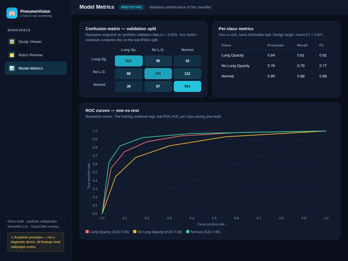
| Evaluation | Per-class precision/recall/F1, confusion matrix, one-vs-rest ROC-AUC — computed by the notebook on your validation split during your build |
| Design target | Macro-F1 approximately 0.80+ on the validation split (target, not a measured claim) |
| Demo | Flask viewer: study view, batch review queue, metrics dashboard |
| Safety status | Academic prototype only — not a diagnostic device, not clinically validated |
Project features
- [RSNA dataset pipeline] DICOM-to-PNG conversion, train/validation/test splits and augmentation (rotations, flips, contrast jitter) for the 26,684-image RSNA set, with the three-class taxonomy preserved.
- [DenseNet-121 classifier] ImageNet-pretrained DenseNet-121 fine-tuned for Normal / No Lung Opacity / Lung Opacity classification, with a training notebook logging loss, per-class metrics and ROC curves every epoch.
- [Grad-CAM explainability] Heatmap overlays generated from the final convolutional block show which lung regions drove each prediction, toggleable in the viewer.
- [Opacity bounding boxes] Predicted boxes with confidence scores localize suspected opacities, mirroring the radiologist annotations in the dataset.
- [Study viewer] Single-study analysis with overlay modes (original, heatmap, boxes, combined), prediction probabilities and DICOM metadata panel.
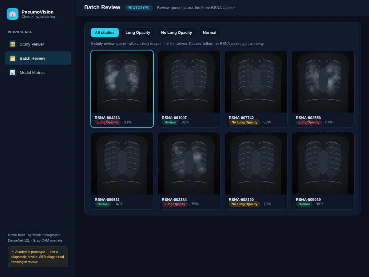
- [Batch review queue] A review grid across all three classes with filtering, so the demo walks through normal and abnormal studies.
- [Metrics dashboard] Confusion matrix, per-class precision/recall/F1 and one-vs-rest ROC curves computed by the notebook on your validation split.
- [Clinical-safety framing] Every screen carries the academic-prototype warning, and the report documents intended use, failure modes and why radiologist oversight is mandatory.
What is included
- Complete source code (dataset pipeline, training, evaluation, Grad-CAM, Flask viewer)
- Jupyter training notebook (buyer-run: preprocessing, fine-tuning, per-epoch metric logging)
- Project report PDF (background, dataset analysis, architecture, evaluation, error analysis, clinical-safety statement)
- PPT presentation for final review
- Viva Q&A preparation document (CNNs, transfer learning, Grad-CAM, ROC-AUC, medical-AI limits)
- Setup guide (environment, downloading the RSNA data from Kaggle, running training and the viewer)
Limitations & prerequisites
- This is an academic prototype and is not intended to replace certified medical equipment, professional diagnosis or clinical decision-making — the report and every demo screen state this explicitly.
- The RSNA labels come from radiology reports with known label noise, and "No Lung Opacity" is a heterogeneous class — the model can inherit that ambiguity, which the error analysis discusses.
- Training covers adult frontal-view radiographs only; performance on pediatric, lateral or portable bedside films is unknown and not claimed.
- No measured accuracy is stated anywhere: the notebook computes per-class metrics on your own validation split during your build, and the report presents those as your build's results against the stated design target.
- The model sees pixels, not patients — it has no access to symptoms, history or lab values that a radiologist uses, so its output is at most a triage signal.
Frequently Asked Questions
Which dataset is used?
The RSNA Pneumonia Detection Challenge (Kaggle, hosted from the 2018 RSNA AI challenge): 26,684 frontal-view chest X-ray images from the NIH Clinical Center, labeled Normal, No Lung Opacity or Lung Opacity, with radiologist-drawn bounding boxes around opacities. The NIH ChestX-ray14 set (112,120 images, 14 labels) is documented as an alternative.
Which model is used?
DenseNet-121 pretrained on ImageNet, fine-tuned with a replaced 3-way classifier head. DenseNet's dense connections suit medical images where subtle texture cues matter and data is limited.
Is the accuracy guaranteed?
No. Nothing is pre-measured — the training notebook logs per-class precision, recall, F1 and ROC-AUC on your validation split every epoch, and the report presents your build's actual numbers. The design target is macro-F1 ≈ 0.80+.
Can it diagnose pneumonia in a real patient?
No — and it must never be presented that way. It is an educational screening prototype. Real diagnosis requires a radiologist with clinical context; the demo and report carry this warning throughout.
What does Grad-CAM show?
A heatmap of which image regions most influenced the predicted class, computed from gradients at the last convolutional layer. It is an explanation aid for the demo and the viva — not proof the model "sees" pathology the way a radiologist does.
Can it work with our college's X-ray images?
Possibly, with caveats: the model expects frontal-view adult radiographs similar to the RSNA distribution. Different machines, pediatric cases or lateral views are out-of-distribution, and the report's limitations section covers exactly this. Suitable for B.E./B.Tech final-year projects in Computer Science, AI/ML and Biomedical-adjacent programs.
Components & software requirements
- Python 3.10, PyTorch (DenseNet-121, transfer learning)
- Torchvision (transforms, augmentation, DataLoaders)
- pydicom + PIL (DICOM conversion and preprocessing)
- scikit-learn (per-class metrics, confusion matrix, ROC-AUC)
- Matplotlib, Seaborn (training curves, heatmaps, ROC plots)
- Flask viewer app (study viewer, batch queue, metrics dashboard)
- RSNA Pneumonia Detection Challenge dataset (Kaggle, 26,684 radiographs)
Delivery information
Built-to-order project. Delivery timeline is shared after order confirmation based on current queue.
Support terms
Complete documentation, setup guide, and viva preparation included. Support for setup and explanation provided.